An Unbiased View of Babyecho
An Unbiased View of Babyecho
Blog Article
Little Known Questions About Babyecho.
Table of ContentsThe Best Strategy To Use For BabyechoBabyecho - QuestionsThe smart Trick of Babyecho That Nobody is DiscussingWhat Does Babyecho Mean?The Only Guide for BabyechoLittle Known Questions About Babyecho.The Greatest Guide To Babyecho

A c-section is surgical procedure in which your child is born through a cut that your doctor makes in your belly and uterus. No matter what an ultrasound reveals, talk to your service provider about the most effective look after you and your child - at home doppler. Last examined: October, 2019
Throughout this check, they will certainly examine the child is growing in the appropriate location, whether there is even more than one infant and they will additionally check your child's advancement up until now. This screening is readily available between 10 14 weeks of pregnancy and is used to assess the opportunities of your baby being birthed with one or even more of these conditions.
Babyecho - Truths
It entails a combined test of an ultrasound check and a blood test. During the scan, the sonographer will certainly gauge the liquid at the back of the baby's neck to figure out 'nuchal translucency' - https://pblc.me/pub/3cb9f10e6009b1. They will then compute the chance of your baby having Down's, Edwards' or Patau's syndrome using your age, the blood test and scan results
During this check, the sonographer checks for architectural and developmental abnormalities in the infant. During this check consultation, you may be offered testings for HIV, syphilis and hepatitis B by an expert midwife. In many cases, a third-trimester scan is suggested by your midwife following the outcomes of previous examinations, previous problems or existing clinical conditions.
The typical 2D ultrasound produces flat and outlined images which can be made use of to see your infant's inner organs and assist spot any type of inner problems. These black and white pictures help the sonographer establish the baby's gestation, growth, heartbeat, development and size. Some pregnant mothers select to have a 3D ultrasound scan due to the fact that they show more of a real-life photo of the baby.
The Best Strategy To Use For Babyecho
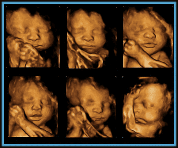
3D ultrasound scans reveal still pictures of your infant's external body as opposed to their insides, so you can see the shape of the baby's facial functions. 4D ultrasound scans are comparable to 3D scans however they reveal a relocating video rather than still images. This captures highlights and darkness better, for that reason creating a clearer image of the child's face and activities.

A is identified throughout this check. A lot of moms and dads choose for this check for.
Babyecho for Beginners
Occasionally a might be called for to get and a clearer photo. This is typically executed and sometimes a might be needed (baby heartbeat doppler). Verify that the baby's heart is existing; To more precisely.
Please see below. It's the same as 19-22 weeks, but some may be or in the and it might to. Typically this is provided if there are such as spina bifida or if parents are keen to understand the earlier. These scans may be done, nonetheless several of the and for this reason, a is needed to This check is done typically at.
Babyecho for Beginners

Additionally, the can be by by an. () The way nearer the is beneficial to. Periodically, an which was previously might be.
The smart Trick of Babyecho That Nobody is Discussing
If, these scans might be to. (of the baby) can additionally be executed. This consists of, along with; This includes, along with (14-20 weeks).
A scan is necessary prior to this examination is done. If you're trying to find, set up an appointment with Dr Sankaran using her. Obstetrics & gynaecology in London.
The Greatest Guide To Babyecho
The test can give valuable information, helping ladies and their health-care service providers take care of and care for the maternity and the unborn child.
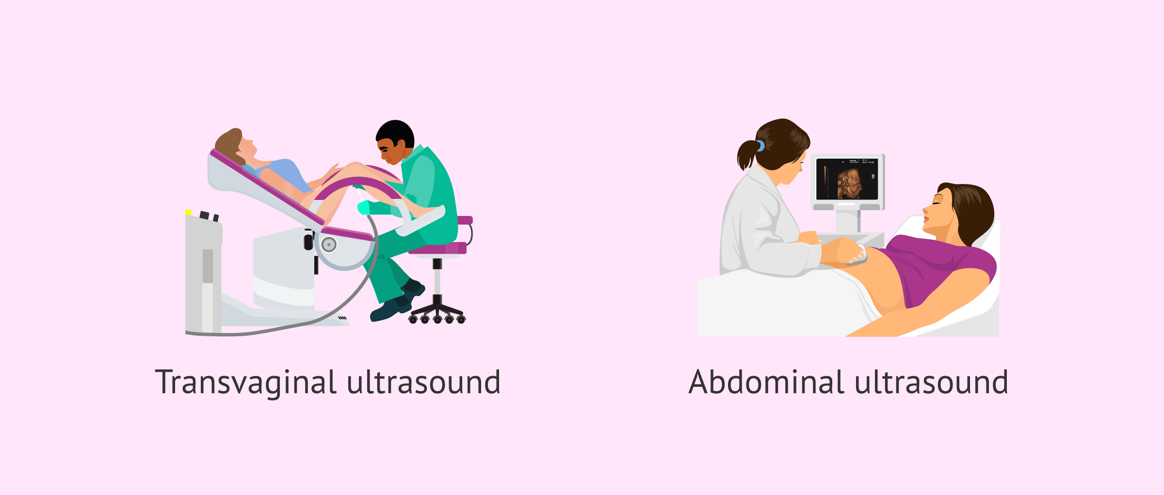
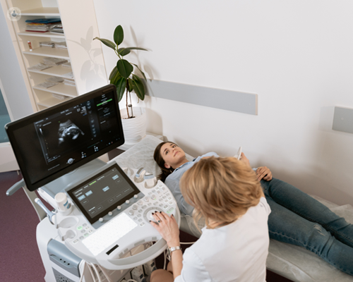
A transducer is inserted right into the vaginal canal and relaxes against the back of the vaginal canal to create a photo. A transvaginal ultrasound produces a sharper image and is frequently made use of in early maternity. Ultrasound machines are about the dimension of a grocery store cart. A TV screen for watching the images is affixed to the maker (https://www.artstation.com/leroyparker5/profile).
Report this page